Detection of Small Events in Environmental Monitoring Culture Media
Automated Incubation and Reading versus Visual Inspection
Laurent Leblanc and Katia Imhoff, R&D, Pharma Quality Control Business Unit, bioMérieux

Abstract
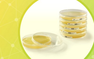
USP chapter <1116> describes Environmental Monitoring (EM) as a key element to ensure control of aseptic processing areas. The quality of the drugs manufactured is directly linked to the capacity to minimize microbial contamination. The only technique to understand and follow the contamination evolution of these critical clean rooms is the usage of specific culture media that can recover the environmental flora. Generally, Petri dishes (plated culture media) are used to monitor air and surfaces and cultivate potential microorganisms found in clean rooms and adjacent areas. The common accepted practice for the examination of Petri dishes consists, after an appropriate incubation (temperatures and time), to enumerate discrete colony forming units (CFU). The microorganisms should then grow into distinct macroscopic colonies. The macroscopic scale describes characteristics a person can directly perceive, without the support of magnifying devices. This means that operators directly observing the Petri dishes should distinguish the presence of microogranisms with the naked eye.
But what limits qualify a macroscopic object? And at what level of size can detection be considered accurate? To answer those questions, a standardized methodology to evaluate the performance of manual reading is presented in this study.
Introduction
EM is one of the main microbiological controls performed by the pharmaceutical industry. This ensures the safety and efficacy of therapies and is a critical control to consider when companies release finished drug products to market. The historical method is extremely manual, variable and error-prone, but it remains the standard procedure used in the industry for hundreds of millions of samples per year.
Indeed, EM trend analysis relies on the accuracy of the results recovered on all Petri dishes used in critical production environments. The analysis is quantitative, the results are expressed in CFU. The visual examination of these plated culture media is realized by qualified operators that must comply with the microbiological best laboratory practices described in the USP chapter <1117>.
In addition to a solid educational background, internal training has to be followed to ensure accurate and reproducible results, although there is no reason to have discrepancies on results between different trained operators. Standardized results are difficult to achieve due to the natural human variation in sight and evaluation. The huge variety of microorganism morphology can complicate detection and enumeration. This is illustrated by numerous FDA 483 forms (12) issued between 2011 and 2021 due to flawed practices during Petri dish inspections; six FDA 483 infractions are attributable to counting errors.
Such observations call into question the accuracy of the results generated during visual inspection of Petri dishes. Therefore, we developed a specific methodology to standardize and measure the quality of detection including challenging events on culture media dedicated to EM.
Download White Paper
Fill out the short form below to download the full white paper.


